Have any questions?
+44 1234 567 890
Without losing their antigenicity or ability to function viable cells of suspensions of any body fluid can be anchored to the adhesion slide and investigated for surface antigens, intracellular antigens, cytochemical functions and morphological properties.
The cells adhere so firmly to the slide that they may be washed without coming off. The extremely hydrophobic mask prevents the cells or reagents from spreading into other wells even if the slide is shaken on a Vortex mixer.
The adhesion slide stops losses of cells, saves time and avoids unnecessary steps of centrifugation.
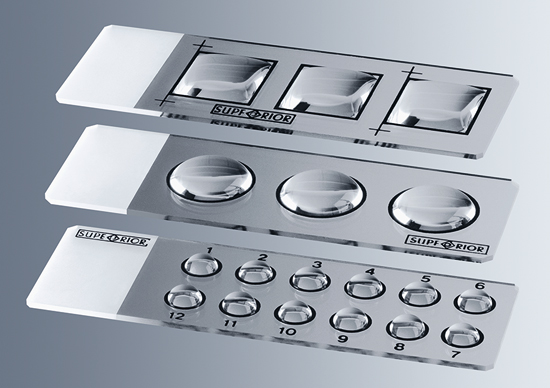
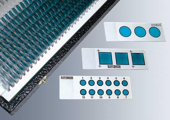
Using a minimum of time and material will result in durable preparations of excellent quality. Up to twelve different tests with different antibodies would be possible on one single slide.
- made of soda lime glass of third hydrolytic class
- in compliance with DIN ISO 8037-1
- dimensions: approx. 76 x 26 mm
- thickness: approx. 1 mm (tol. ± 0.05 mm)
- with 90° ground edges
- with clipped corners
- with silky frosted marking area of approx. 15 mm on one side
- in storing boxes of 50 or 100 pieces
- not registered for in vitro diagnostic use within the EU
| Cat. No. | Type | Unit |
|---|---|---|
| 0900000 | 12 x 5 mm Ø | 50 |
| 0900100 | 12 x 5 mm Ø | 100 |
| 0901000 | 3 x 15 mm Ø | 50 |
| 0901100 | 3 x 15 mm Ø | 100 |
| 0906000 | 3 square wells 15x15 mm and 4 register marks | 50 |
| 0906100 | 3 square wells 15x15 mm and 4 register marks | 100 |
Instructions for PAP and APAAP slide assay can be downloaded here: Application of the Adhesion slide
Preparations on the adhesion slide
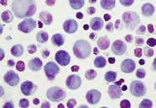
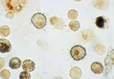
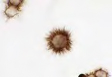
(left to right)
(1) Hematoxylin staining
(2) Immunocytologic double staining for CD36 (dark blue ring indicating membrane staining) and Interleukin 8 (brown dots within the cells). Note the hairy appearance of the dendritic cell membrane, which is uniquely preserved after fixation with glutaraldehyde. (Immunostaining with sequential ABC technique using 4-CN as substrate for CD36 development and DAB for IL-8; D. Behringer, Zellmarkerlabor, University Hospital of Freiburg)
(3) D20 reactive malignant B-lymphocytes isolated from the cerebrospinal fluid of a patient with B-cell lymphoma thus indicating meningeal spread of the disease. Note the typical hairy appearance of these cells. (Immunostaining with immunoperoxidase technique using DAB for CD20 development; fixation: 0.04% glutaraldehyde; D. Behringer, Zellmarkerlabor, University Hospital of Freiburg)